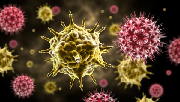
A team from Stanford University utilized cutting-edge microscopy to delve into the intricacies of SARS-CoV-2 replication within cells, potentially revolutionizing drug development. They captured high-resolution images of the virus’s RNA and replication structures forming around the cell’s nucleus.
The study explained the molecular activity of the virus inside host cells in detail. Viruses take over cells, turning them into virus factories with particular replication sites. Here, the viral RNA duplicates until enough is made to infect new cells.
Stanford scientists focused on seeing this replication step in sharp detail. They labeled the viral RNA and proteins with fluorescent colors. To avoid fuzzy images, they used a chemical to randomly make the fluorescence blink on and off. This helped them pinpoint the exact locations of the molecules.
The researchers used lasers, robust microscopes, and a fast camera to capture detailed images of blinking molecules. Combining these images created clear photos showing the cells‘ viral RNA and replication structures.
With a 10-nanometer resolution, the images reveal the most detailed view of the virus replicating inside a cell. The magenta RNA forms clumps around the cell’s nucleus, creating a sizeable repeating pattern.
The clusters reveal how the virus avoids the cell’s defenses. They gather inside a membrane to hide from the rest of the cell. Unlike electron microscopes, the new imaging technique uses blinking fluorescent labels to pinpoint virus components in a cell. This method shows nanoscale details invisible in traditional biochemical assays.
Seeing how the virus infects cells can improve medicine. Observing how different viruses take over cells may explain why some cause mild symptoms while others are deadly. Super-resolution microscopy can aid drug development. This nanoscale structure of the replication organelles can provide new therapeutic targets.
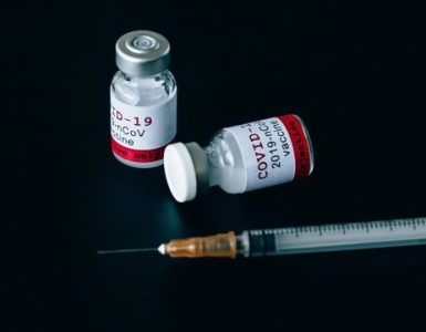
The team plans to repeat the experiment with drugs like Paxlovid or remdesivir to see if they can suppress viral replication, which would indicate the drug’s effectiveness. Stanley Qi, Stanford associate professor of bioengineering in the Schools of Engineering and Medicine and co-senior author of the paper, said, “We have not seen COVID infecting cells at this high resolution and known what we are looking at before. Knowing what you are looking at with this high resolution over time is fundamentally helpful to virology and future virus research, including antiviral drug development.”
Source: Tech Explorist